
Bone fractures are common among active individuals, but not every fracture is the same. Doctors use imaging technology to assess injury and diagnosis. An X-ray is common, providing a look inside the body. This technology enables providers to evaluate damage. When you visit a clinic for an injuries, often the first step is imaging. X-rays pass radiation through the body, and different tissues absorb it at varying rates, allowing doctors to visualize bones and joints.
The Mechanics of X-Ray Imaging
The process begins with positioning the injured area so the X-ray beam can pass through it. A technician places you between the X-ray machine and a digital detector, making sure to align you. You must stay still, or the image will blur and become unusable, which can delay the diagnosis. This stillness is so the resulting picture is sharp, allowing doctors to see even small fractures.
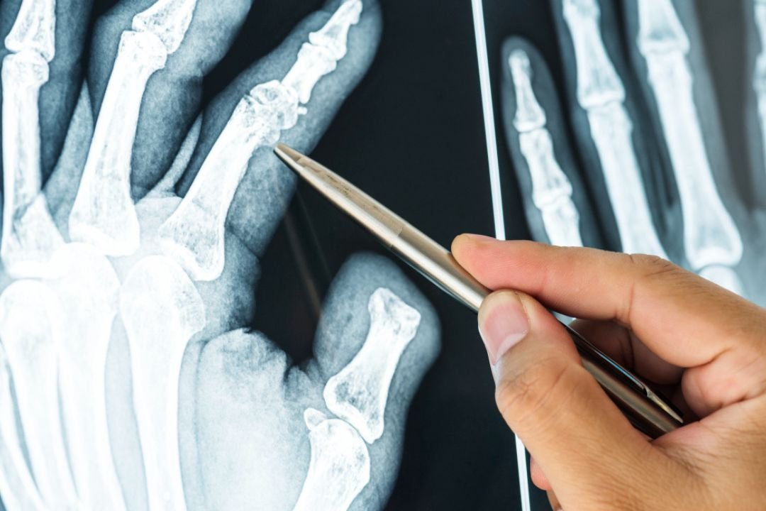
The machine sends a quick burst of radiation through the limb, and the detector captures the energy that passes through. After the energy is captured, a computer processes a digital image. Dense structures block energy, creating the white shape of bones, and in contrast, soft tissues appear darker. Fractures appear as dark lines because the beam passes through the break, making them distinguishable. Physicians analyze these images to determine the breaks severity, and they look for alignment issues or fragments. If the injury is complex, they might order an MRI for more detail, but an X-ray usually provides enough information.
Identifying Different Types of Fractures
Not all bone breaks look the same on an image. Some are simple cracks, while others involve multiple fragments and are more complex. A doctor might see a hairline fracture or a complete break. The treatment plan depends on what is seen in the image. Stress fractures, which are tiny cracks from overuse, often cause foot or ankle pain. These fractures can be hard to see at first, so follow-up images may be needed. Rest is usually the main treatment, but sometimes therapy is used.
Trauma can cause more severe breaks that displace the bone, which makes healing more challenging. In these cases, the bone ends do not align, and surgery may be required to restore normal function. Specialists use X-rays to carefully plan these surgeries, and they rely on detailed images to make precise decisions. They check that the bone is set correctly, so patients can regain mobility as quickly as possible.
Guiding Treatment and Recovery
Once a diagnosis is made, the focus shifts to recovery, and an individualized plan is created. The images guide the doctor in choosing a cast, brace, or surgical pin, depending on the injury type and severity. You will likely need follow-up appointments, and new images will track your progress so any issues may be caught. This monitoring checks that the bone is healing, and adjustments can be made.
For issues like elbow or shoulder pain, the approach might differ because different joints unique. If the joint is involved, the doctor also checks for cartilage damage, which can complicate recovery. They might suggest PRP therapy, or prescribe injections for relief, although physical therapy may also be an option. The goal is always to restore function and reduce pain, regardless of the complexity.
Schedule Your Consultation Today
Your provider is here to help you heal and support you throughout your recovery. If you are experiencing pain, prompt diagnosis can help, since early intervention improves outcomes. Your team uses advanced imaging to identify fractures, and create a personalized treatment plan for you. Schedule an appointment for your injury assessment, and start toward a full recovery.
Leave a Reply